 |
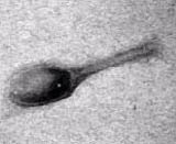
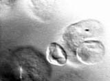
MOVIE 1-This movie illustrates
the tilt of a T4 bacteriophage. Notice the
depression of the virus head asthe movie plays. Without any specimen motion,
it is difficult to discern the depression. (TEM)
Courtesy Sheila Warren.
File Size- 81KB |
|
|
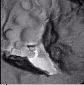 |
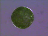
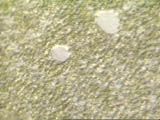
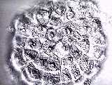
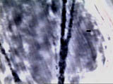
MOVIE 2-Saccharomyces
cerevisiae.The motion of a freeze-fractured
yeast cell is depicted in the movie above. Before the movie begins,
one has no idea of depth perception; however, when the movie starts, one
will immediately use his/her depth perception cues and be able to easily
discern convex structures from concave structure. Notice the protruding
portion of a membrane in the lower left corner. This is really a prominent
tear of the freeze fracture replica protruding 90 degrees above the replica
surface. The large, convex structure in the is the nucleus, with its prominent
nuclear pores. There should be several concave depressions
on the nucleus. This series of speicmen tilts was made using a eucentric
goniometer stages on our Philips 420 transmission electron microscope.
The stage can be tilted +- 60 degrees. Compare this movie with the 3-d
images at:
Courtesy R. Malcolm Brown, Jr. and Sheila Warren.
File Size- 206KB |
|
|
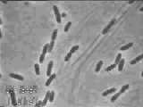 |
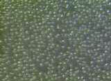
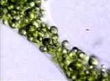
MOVIE 3-Acetobacter
xylinum synthesizing cellulose. In this video clip it is useful
to observe the backward and forward motion of the Acetobacter cells.
This movement is known as reversals. Although reversals are not completely
understood, it is known that the motion is caused by cellulose
synthesis rather than flagella. Recent research suggests that buildup
of strain during the crystallization of cellulose may be responsible for
these reversals.
The linear elongation rate of cellulose from the surface of the bacterium
can be used to calculate the rate of cellulose deposition. For example,
a typical elongation rate of 2 um per min means that more than 10 8
glucose molecules are incorporated into cellulose per hour per bacterial
cell. A static culture with a surface area of one acre could produce more
than 20,000 pounds of cellulose per year (compare with 600 pounds per acre
per year as a typcial yield for a bale of cotton per acre).For information
on how to culture Acetobacter, click HERE
Time-lapse video by Martin Spiess, movie conversion courtesy Sheila
Warren.
File Size- 501KB |
|
|
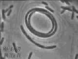 |
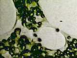
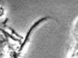
MOVIE 4-Acetobacter
xylinum synthesizing cellulose. In this video clip notice the uncoiling
of the Acetobacter cells. Like the reversals, the uncoiling motion
is caused by cellulose synthesis. When the Acetobacter uncoils, bundles
of cellulose ribbons can be seen in the background as dark grey filaments.
These are sub-micron nano-structures. Notice that the Acetobacter cells
continue to synthesize multiple cellulose ribbons even though they are
in attached chains and incompletely separated.
Time-lapse video by Martin Spiess, movie conversion courtesy
Sheila Warren.
File Size- 1,245KB |
|
|
 |
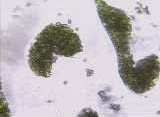
 MOVIE
5-Boergesenia forbesii synthesizing
cellulose. This is a time-lapse video taken at 3 hours post-wounding and
continuing for a period of 5 hours at one frame every 40 seconds (435 frames
total). These images were taken under a polarized light microscope with
an attached color CCD camera (Optronix Camera) coupled to a Panasonic optical
disk recorder. The bright ring which forms around the Boergesenia
aplanospore is due to the birefringence of the cellulose wall which is
being synthesized around the newly formed cell. It is possible to correlate
the quanity of cellulose being synthesized with the appearance and change
in the birefringence. This is the first example of direct visualization
of cellulose synthesis in a cell wall of a eukaryotic organism. MOVIE
5-Boergesenia forbesii synthesizing
cellulose. This is a time-lapse video taken at 3 hours post-wounding and
continuing for a period of 5 hours at one frame every 40 seconds (435 frames
total). These images were taken under a polarized light microscope with
an attached color CCD camera (Optronix Camera) coupled to a Panasonic optical
disk recorder. The bright ring which forms around the Boergesenia
aplanospore is due to the birefringence of the cellulose wall which is
being synthesized around the newly formed cell. It is possible to correlate
the quanity of cellulose being synthesized with the appearance and change
in the birefringence. This is the first example of direct visualization
of cellulose synthesis in a cell wall of a eukaryotic organism.
Time lapse video by Andrew Bowling, movie conversion courtesy Sheila
Warren.
File Size- 394KB |
|
|
 |
MOVIE
6-Boergesenia forbesii -completeprotoplast
formation. Note the motion of the protoplast during the budding
phase in which individual segments are being formed. You will be greatly
surprised at the final outcome of this movie. It will look NOTHING
like the beginning!
Courtesy Andrew Bowling.
File Size- 307KB |
|
|
 |
MOVIE
7-Boergesenia forbesii-
higher magnification view of an early stage of protoplast formation. Note
that the final separation of protoplasm occurs through the formation of
a narrow bridge, ultimately separating the protoplast. Courtesy
Andrew Bowling.
File Size- 254KB |
|
|
 |
MOVIE
8-Boergesenia forbesii-
a later stage of protoplast formation giving rise to spherical protoplasts.
Courtesy
Andrew Bowling.
File Size- 100KB |
|
|
 |
MOVIE
9-Boergesenia forbesii-
sequence from initial cleavage to beginning separation of protoplasts.
This takes place within 90 min after wounding. Courtesy Andrew
Bowling.
File Size- 340KB |
|
|
 |
MOVIE
10-Boergesenia forbesii-
Excellent,complete sequence from initial cleavage to separation and formation
of spherical protoplasts. Courtesy Andrew Bowling.
File Size- 539KB |
|
|
 |
MOVIE
11-Boergesenia forbesii-another
higher magnification view showing details of the migration of protoplasm
to from the initial stages of segmentation. Courtesy Andrew
Bowling.
File Size- 163KB |
|
|
 |
MOVIE
12-Acetobacter cellulose exposed to cellulase
(CBH I). When a complete cellulase mixture
is added to the cellulose suspension (eg, CBHI, CBHII, endoglucanase, b-glucosidase,
etc), a remarkable twisting motion is initiated. No living cells are present.
This motion is believed to be the result of strain released as the
microfibrils are being degraded by the cellulase. Experiments are in progress
to determine the specific site and nature of this interesting dynamic motion.
At present we do not know if only one or several cellulases are required
for the induction of this motion.
Please examine this video carefully and run it
several times. Look first at the large multistranded cable of cellulose
microfibrils. The rotation is counter-clockwise. Next, look at the two
cellulose ribbon bundles which are perpendicular to this large strand.
At first, they are intact, but one of them literally "dissolves" as the
cellulase action continues. Then on another try, focus your attention on
the material at the right-hand side of the movie. You will see it dissolve
also! To our knowledge, this is the first dynamic account of cellulase
action. Video scenes, courtesy Robert Lindstrom and Andrew Bowling.
Cellulase and bacterial cellulose, courtesty Yoshihiko Amano.
File Size- 146KB |
|
|
 |
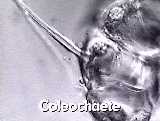
MOVIE
13- Coleochaete scutata hair
cell rotation. In this unusual dynamic time lapse move made by RM Brown
when he was in Germany in 1969, an algal colony of Coleochaete
cells is observed in which the basal cells oscillate while the hair cells
undergo a continuous rotation. This rotation is believed to be the result
of a directed secretory process through the hair cell protusions. Virtually
nothing is known about the mechanism for such motion. Permission granted
from the Institute fur den Wissenschaftlichen
Film Gottingen, Germany.
  
By the way, the IWF is one of the world's finest
film institutes.
When in Germany, be sure to visit this fantastic research lab and search
through the archives of thousands of scientific films. Many of these are
now available on video for teaching and research. If you would like to
review details of these movies I made, go to my IWF Movie Detail Page-Click
HERE.
File Size- 406KB |
|
|
 |
MOVIE
14-Coleochaete scutata hair
cell rotation. This scene shows in detail an oblique view of a single hair
cell rotating. The single chloroplast is quite obvious as it spins around
in the hair cell's basal region. Permission granted from the Institute
fur den Wissenschaftlichen Film Gottingen, Germany.
File Size- 542KB |
|
|
 |
MOVIE
15-Prymnesium parvum Golgi
apparatus dynamically imaged! To my knowledge,
this is the only light microscopy imaging of a functioning Golgi apparatus
in a living cell. Look at the arrow overlay which soon appears. In what
looks like contractile vacules, the individual Golgi cisternae are imaged,
forming initially as flattened sacs, then enlarging as they move
to the cell surface where they deposit scales via exocytosis . The scales
are a modified cell wall. Notice the background for this webpage is is
a Golgi-derived cellulosic scale from the alga, Pleurochrysis scherfellii.
Permission granted from the Institute fur
den Wissenschaftlichen Film Gottingen, Germany. Coming
soon! details of movies 15 and 16!
File Size- 504KB |
|
|
 |
MOVIE
16-Prymnesium parvum Golgi
apparatus dynamically imaged! This second movie show the Golgi secretion
of scales from an oblique view, with the scales being secreted perpendicular
to the plane of the image. Permission granted from the Institute
fur den Wissenschaftlichen Film Gottingen, Germany.
File Size- 582KB |
|
|
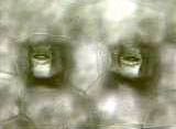 |
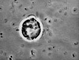
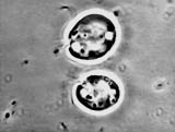
MOVIE
17-Tradescantia-two
stomata with guard cells, resembling two eyes! This short video is a thru-focus
on the lower surface of the leaf.
Video scenes, courtesy R. Malcolm Brown, Jr.
Tradescantia, courtesy, Robert Jackson.
File Size- 151KB |
|
|
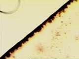 |
MOVIE
18-Linear acetylenic carbon-This
video begins with a boundary of linear acetylenic carbon (LAC) dried from
an organic solvent onto a microscope slide. In the scene, water is added
(bubble at the top left) which mixes with the LAC forming strand-like threads
resulting in the viscous alignment of the carbon chains which are
comprised of at least several hundred carbon atoms each. LAC courtesy of
Richard Lagow, Department of Chemistry, UT-Austin (see Lagow et al. Science267:
p 362 1995).
Video scenes, courtesy R. Malcolm Brown, Jr.
and Judith Sharp.
File Size- 816KB |
|
|
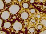 |
MOVIE
19-Linear acetylenic carbon-This
video begins with a boundary of linear acetylenic carbon (LAC) dried from
an organic solvent onto a microscope slide. In the scene, water is added
which mixes with the LAC forming thread like structures within each of
the clear areas resulting in the viscous alignment of the carbon
chains. LAC courtesy of Richard Lagow, Department of Chemistry, UT-Austin
(see Lagow et al. Science 267: p 362 (1995)
Video scenes, courtesy Shelley Behlen.
File Size- 118KB |
|
|
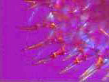 |
MOVIE
20-Tradescantia
hair cells imaged in polarized light. This video shows the first order
red compensator addition colors as these birefringent hair cells are rotated
360 degrees. The birefringence due to the molecular order of cellulose
microfibrils in the wall, is indicative of a precise, ordered deposition
of cellulose within the wall.
Video scene, courtesy R. Malcolm Brown, Jr.
File Size- 120KB |
|
|
 |
MOVIE
21-Paramylon- These are crystalline grains
of B-1,3 glucan from Euglena.This material also is known as "curdlan".
The video shows dissolution the curdalan upon being treated with 1.0M NaOH.
Original materials supplied by Iain Cheeseman-
Click
HERE
to learn more. Video scene, courtesy R. Malcolm Brown, Jr.
File Size- 82KB |
|
|
 |
MOVIE
22-Tradescantia
staminal hair cells demonstrating cytoplasmic straming . There are
several excellent scenes in this video.
Video scene, courtesy R. Malcolm Brown, Jr.
File Size- 376KB |
|
|
|
|