|

| |
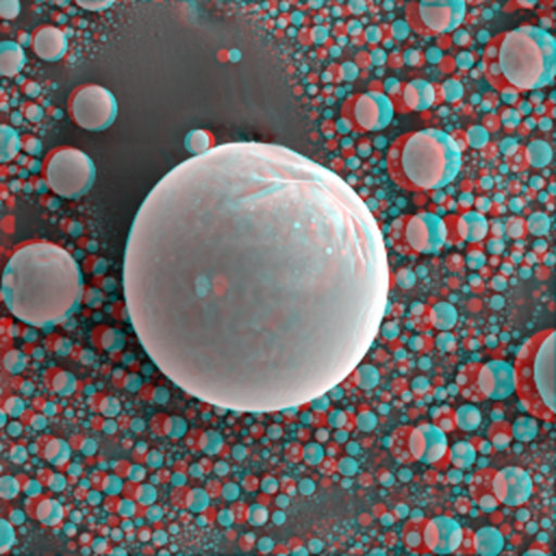
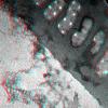
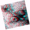
This is an image of polystyrene beads imaged with a scanning electron microscope. It is an excellent test sample for stereo imaging
|
|---|
| 
| |
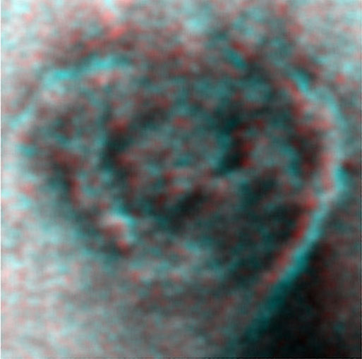
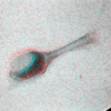
The image on the left shows mucilage (or perhaps other diatom debris) associated with the frustule of a marine diatom, Stauroneis sp. Due to the thickness of the specimen, it is difficult to discern the point of origin of the extracellular material. (TEM)
|
|---|
|
| |
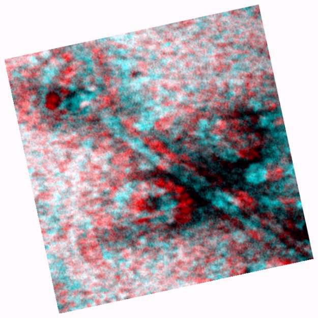
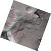
This is an excellent 3-D example of extracellular matrix being extruded from the raphe of Stauroneis sp.. On the left, curved filaments are found at the end of a mucilage stream from the diatom. It is unclear whether these filaments may be involved with diatom adhesion to surfaces such as a Formvar-coated copper grid or glass slide. (TEM)
|
|---|
|
| |
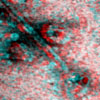
A T4 bacteriophage is depicted on the left. The three-dimensional appearance of this image is not as obvious due to the angle of this particular specimen's tilt. For this specimen, the three-dimensional qualities of the head and tail are much better represented by an animation of the tilt. (TEM)
|
|---|
|
| |
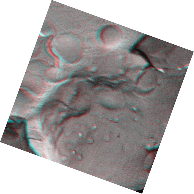
The three-dimensional appearance of a freeze-fractured yeast cell is shown in this image. The convex structure the middle foreground is the nucleus, covered withsmall pits known as nuclear pores (these are the structures through which mRNA and other nuclear components pass through en route to the cytoplasm. Also, notice other cytoplasmic vacuoles in the background. A piece of replica upturned 90 degrees is obviously projecting perpendicularly to the fracture surface. This example demonstrates well the usefulness of the anaglyph in assisting in the interpretation of three dimensional nanostructure (TEM)
There is also a movie of this image demonstating continuous tilting of the specimen.(Go to the movie section)
|
|---|
|
| |
This image contains a cellulose microfibril labelled with a cellobiohydrolase/gobl comdex. Attached to the microfibril are cellulose synthase enzyme particles which are roducing cellulose (glucon chains) from a VDP glucose monomomer.
|
|---|
|
| |
Cellulose synthsizing complex isolated from Acetobacter xylinum. Two complexes stratle a presynthesized cellulose microfibril. They are held together by newly synthesized glucan chains. Colloidal gold-CBHI is a specific probe for cellulose. Courtesy Tara Spires
|
|---|
|

| |
This is an anaglyph of graphite at a total magnification of 19,440,000X. The 3.35Ĺ spacing between layers in the graphite lattice is clearly shown, and the red-blue separation allows perception of depth in the figure. This anaglyph was made using two images of the same piece of graphite taken at 5° tilt separation from each other. Both images were transformed into a red-green-blue pixellation scale. The red pixels were subtracted from one, the blue from the other, and the images were superimposed. Courtesy Tara Spires
|
|---|
|

| |
Another stereo image of graphite. The graphite images were prepared by tilting the specimen at very high manginfication and digitizing two seperate images which were then processed for the anaglyph. Courtesy Tara Spires
|
|---|