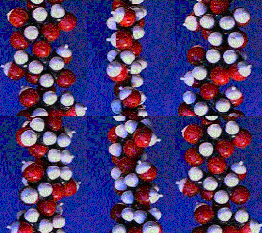
Space filling model of a glucan chain of cellulose. This is a large file, but the model should ROTATE!

Anaglyphs
![]() The construction of anaglyph images is useful, in some cases, for understanding the three-dimensional organization of biological specimens that are observed with the transmission electron microscope (TEM).
The construction of anaglyph images is useful, in some cases, for understanding the three-dimensional organization of biological specimens that are observed with the transmission electron microscope (TEM).
This summer, Sheila Warren embarked on a program to study techniques and methodology which might be useful in the 3-D imaging of structures using the transmission electron microscope (TEM). Presented below will be a NEW WORLD of NANOSTRUCTURE observed in a rather unique way.
When studying specimens with the TEM, one of the limitations involves seeing only one perspective at a time. Consequently, it is sometimes difficult to visualize structures in three dimensions. To overcome this limitation, specimens were tilted and a separate images were captured for every tilt increment. Two images of the same specimen at different tilts were then selected to form a stereoimage. Taken a step further, the images composing the stereoimage were assigned shades of blue or red, and then they were combined to form a 3-d image that is viewed with red/blue glasses. Following this passage are images that illustrate the usefulness of anaglyphs in examining biological specimens. Don't forget your 3-d (red/blue) glasses! If you do not have these, a good source of contact is Reel 3-D Enterprises, Inc. P.O. Box 2368, Culver City, California 90231 Tel (310) 837-2368. When wearing these glasses, the blue lens should be in front of the right eye. Now go fetch some really interesting STUFF!