in a New Supermolecular Structure of Cellulose
Tetsuo Kondo*1, Eiji Togawa1 and R. Malcolm Brown Jr.2
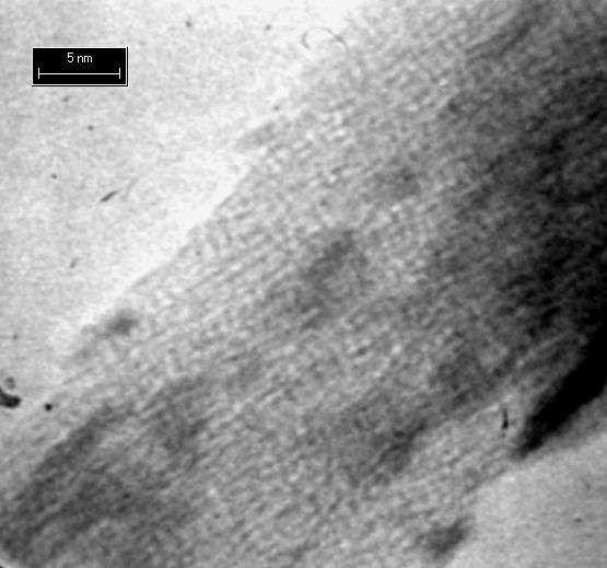
Fig.1 High resolution TEM image of single b-glucan chains in ordered, non-crystalline regions of quasi-tactic cellulose. The TEM sample specimen was also prepared from the same cellulose/DMAc/LiCl solution as previously demonstrated in (1) but diluted by 100 times with water-free DMAc (1.0x10⁻² wt%). After the solution was poured over a 3.0mm electron microscope grid coated with Formvar, the grid was incubated under a saturated water vapor overnight. It was washed with distilled water, and then the sample on the grid was negatively stained with 2% uranyl acetate. Interaction with uranyl acetate appears to protect samples from electron beam damage.
Scale bar indicates 5 nm length.
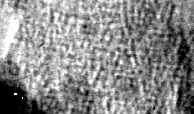
Fig. 2. Higher magnification view of Fig 1 above. Note the 0.24nm crystalline lattice of uranyl acetate is clearly resolved. Individual glucan chains are clearly decorated by the uranyl acetate. Note the order of the glucan chains; however, they are not crystalline.
Scale bar indicates 2 nm length.